
last update on 2006.8.15
Staffs
- Masao Iwase (Instructor)
- Ryouhei Ishii (Instructor)
- Kiyotake Takahashi (Resident)
- Hidetoshi Takahashi (Resident)
- Takayuki Nakahachi (Post-doctoral fellow)
- Ryuji Sekiyama (Graduate student)
- Eiko Honaga (Graduate student)
- Ryu Kurimoto (Graduate student)
- Leonides Canuet (Graduate student)
- Kouji Ikezawa (Graduate student)
- Michiyo Azechi (Graduate student)
Our research themes
In our laboratory, we are investigating schizophrenia, depression, autism spectrum disorders, dementia and epilepsy using cognitive behavioral methods and neurophysiological methods like Magnetoencephalography (MEG), electroencephalograhy (EEG), near-infrared spectroscopy (NIRS), positron emission tomography (PET) and transcranial magnetic stimulation (TMS). Our research themes are pathophysiology and therapeutic methods of these neuropsychiatric disorders.
1. PET
A collaborative PET study of human laughter revealed that orbitofrontal cortex was associated with mirth experience and that supplementary motor area and basal ganglia were associated with facial expression of laughter.
2. MEG
We indicated that frontal medial theta wave (Fmθ) generated from medial frontal lobe and that there were theta activities in the lateral parts of frontal lobe.
Transient activities of dorsolateral prefrontal cortices during Stroop task were detected using MEG.
The neural activity of language associated area during last-and-first word game task (shiritori) were visualized. This task was applied to the patients with schizophrenia and contributed to the elucidation of its pathophysiology.
Neural activities of temporal lobe during auditory hallucination of patients with schizophrenia were detected using MEG.
We estimated the loci of neural activities of epileptic patients with auditory hallucination and compared pathophysiology of epilepsy with that of schizophrenia.
3. EEG
Statistical and mathematical analysis of EEG data revealed regional cerebral activities during higher cognitive functions and abnormalities caused by aging and dementia.
4. NIRS
A collaborative study revealed the regional changes of cerebral blood flow during Fmθ generation.
5. rTMS
A basic research on the therapy of thalamic pain is ongoing. We are also preparing rTMS studies of depression and schizophrenia.
6. Psychoneuroimmunology
The elevation of natural killer cell activity by laughter was indicated using a crossover study.
7. Cognitive impairments in schizophrenia and autism spectrum disorders
Using PC-based cognitive task, the association of cognitive impairments with clinical features were investigated in schizophrenia and autism spectrum disorders.
Neural basis of social malfunctioning in Asperger's disorder is now investigated using neurophysiological methods.
8. Our collaborators
- Department of Physiology, Osaka City University Graduate School of Medicine.
- Kansai University of Social Welfare
- Wakayama Medical University
- Gifu University
- University of Toronto
- Department of Neurosurgery, Osaka Univesity
- Department of Radiology, Osaka University
- Department of Anesthesiology, Osaka University
- Department of Physiology (Molecular Physiology), Osaka University
Figure
This figure indicates the brain regions where regional cerebral blood flow and the amount of laughter were highly correlated. The more the amount of laughter, the more neural activities in supplementary motor area (A) and left putamen (C). In the graph B and D indicates, the amount of laughter and rCBF change were plotted in the horizontal and vertical axis, respectively.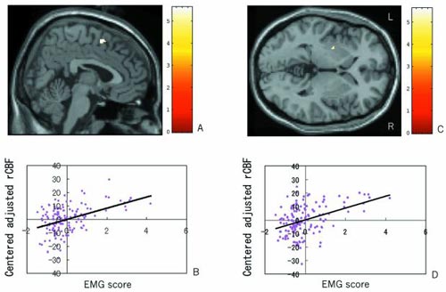
This figure shows the activated areas during laughter compared with voluntary face movement. In these activated areas, orbitofrontal area (D) and medial prefrontal area (E) are considered involved in the mirthful experience of laughter. The activated areas in occipital, occipitotemporal and anteriotemporal areas are considered the cognition of humorous visual stimuli.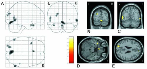
Articles
- Discrepancy of performance among working memory-related tasks in autism spectrum disorders was caused by task characteristics, apart from working memory, which could interfere with task execution. Nakahachi T, Iwase M, Takahashi H, Honaga E, Sekiyama R, Ukai S, Ishii R, Ishigami W, Kajimoto O, Yamashita K, Hashimoto R, Tanii H, Shimizu A, Takeda M. Psychiatry and Clin Neurosci, 60: 312-318, 2006
- Information processing flow and neural activations in the dorsolateral prefrontal cortex in the Stroop task in schizophrenic patients. A spatially filtered MEG analysis with high temporal and spatial resolution. Kawaguchi S, Ukai S, Shinosaki K, Ishii R, Yamamoto M, Ogawa A, Mizuno-Matsumoto Y, Fujita N, Yoshimine T, Takeda M. Neuropsychobiology. 2005;51(4):191-203.
- Spatial working memory deficit correlates with disorganization symptoms and social functioning in schizophrenia. Takahashi H, Iwase M, Nakahachi T, Sekiyama R, Tabushi K, Kajimoto O, Shimizu A, Takeda M. Psychiatry and Clin Neurosci, 59: 453-460, 2005
- Slow repetitive transcranial magnetic stimulation increases somatosensory high-frequency oscillations in humans. Ogawa A, Ukai S, Shinosaki K, Yamamoto M, Kawaguchi S, Ishii R, Takeda M. Neurosci Lett. 2004 Apr 1;358(3):193-6.
- Interictal spikes in the fusiform and Inferior temporal gyri of an epileptic patient with colored elementary visual auras - a 5-year longitudinal MEG ECD study -. Shunsuke Kawaguchi, Kazuhiro Shinosaki, Satoshi Ukai, Ryouhei Ishii, Masakiyo Yamamoto, Asao Ogawa, Norihiko Fujita, Toshiki Yoshimine and Masatoshi Takeda. Neuroreport (2003), 14(4): 637-640.
- Current Source Density Distribution of Sleep Spindles in Humans as found by Synthetic Aperture Magnetometry. Ryouhei Ishii, Rainer Dziewas, Wilkin Chau, Peter Soros, Hidehiko Okamoto, Atsuko Gunji, Christo Pantev. Neuroscience Letter (2003), 340 25-28.
- Neural substrates of human facial expression of pleasant emotion induced by comic films: a PET study. Iwase M, Ouchi Y, Okada H, Yokoyama C, Nobezawa S, Yoshikawa E, Tsukada H, Takeda M, Yamashita K, Takeda M, Yamaguti K, Kuratsune H, Shimizu A and Watanabe Y. NeuroImage 17: 758-768, 2002
- Parallel Distributed Processing Neuroimaging in the Stroop task using Spatially Filtered MEG Analysis. Satoshi Ukai, Kazuhiro Shinosaki, Ryouhei Ishii, Asao Ogawa, Yuko Mizuno-Matsumoto, Tsuyoshi Inouye, Norio Hirabuki, Toshiki Yoshimine, Stephen E. Robinson, Masatoshi Takeda. Neuroscience Letter, 2002, 334:9-12.
- The elevation of natural killer cell activity induced by laughter in a crossover designed study. Takahashi K, Iwase M, Yamashita K, Tatsumoto Y, Ue H, Kuratsune H, Shimizu A, Takeda M, Int. J. Mol. Med. 8: 645-650, 2001
- Medial Prefrontal Cortex Generates Frontal Midline Theta Rhythm. Ishii, R., Shinosaki, K., Ukai, S., Inouye, T., Ishihara, T., Yoshimine, T., Hirabuki, N., Asada, H., Kihara, T., Robinson, S.E., Takeda, M. NeuroReport. 10(4) 675-679, 1999.