|
|
|
|
HOME > Programmed research projects > Programmed research project 04
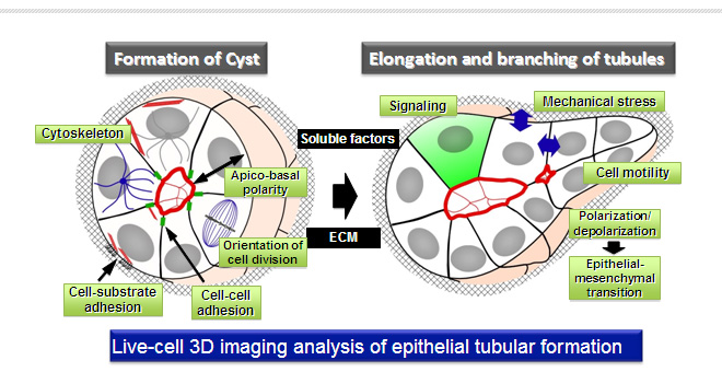
 Programmed research project 04Live-cell 3D imaging analysis of the dynamics of epithelial cells and functional molecules in epithelial tubular formation
Purpose of the Research ProjectEpithelial tissues basically consist of tubular structures of polarized epithelial-cell sheets. Various extracellular and intracellular signals spatio-temporally regulate dynamics of cytoskeletons and cell polarity and adhesion for the morphogenesis and maintenance of epithelial tubules. In this project, using 3D live-cell imaging analysis, we challenge to visualize dynamics of cell architectures in connection with cell adhesion, morphology, migration, polarization/depolarization and division during epithelial tubular formation, and elucidate the molecular mechanisms of spatio-temporal control of tubule formation, focusing on the signaling of Rho GTPases. We also challenge to elucidate the role and signaling mechanisms of mechanical forces in the development of epithelial tubules. Based on the imaging analysis, we would like to clarify the common and tissue-specific mechanisms of actin cytoskeletal remodeling for the maintenance and development of epithelial tissues. Content of the Research ProjectIn this project, we focus on the functional roles of the Rho family GTPases signaling, including their upstream regulators and downstream effectors. To analyze the molecular mechanisms of epithelial tubular formation, we will visualize changes in actin cytoskeleton, cell adhesion, cell polarity and orientation of cell division in 3D-cultured epithelial cells and organ cultures. We will also visualize changes in the activities of signaling molecules and the forces on the cell-cell and cell-substrate junctions using the fluorescent probes. We are launching a plan to develop the fluorescent probes visualizing changes in cytoskeletons, polarity, and mechanical forces. Furthermore, we will seek for the functions of key molecules in tubule formation by the combination of imaging and biochemical analyses. We identified some of the Rho-GEFs involved in matrix stiffness-induced cell transformation of 3D-cultured mammary epithelial MCF10A cells. We are going to analyze the role of the Rho-GEFs in the exchange of cell polarization and depolarization, which importantly contributes to the tubular formation. Expected Research Achievements and Scientific SignificanceIn this project, we develop the high-resolution live-cell imaging system in 3D-cultured epithelial cells. Our research will allow us to visualize the patterns of dynamics of actin cytoskeletal remodeling, cell adhesion and cell polarity in each step of epithelial tubular formation. Progress in imaging techniques will serve to understand the functional roles of key signaling molecules and mechanical forces in epithelial tubular formation. The outcome will provide novel insights into the molecular mechanisms of the maintenance and development of epithelial tubular tissues. Our researches will contribute to a better understanding of organogenesis and tumorigenesis and the progress of regenerative medicine.
|