|
|
|
|
HOME > Proposed research projects > 2012-2013: Proposed research projects 15
 2012-2013: Proposed research project 15Development of a novel imaging system in branching morphogenesis of the lung using optical coherence tomography
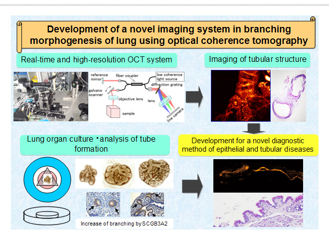
Purpose of the Research ProjectOptical coherence tomography (OCT) is an emerging optical imaging modality in biomedical optics and medicine. OCT provides high-resolution, cross-sectional images of the internal microstructure in biological tissues by measuring echos of backscattered light. Tissue pathology can be imaged in situ and in real time with resolution of 1-15 µm, one to two orders of magnitude finer than that of conventional ultrasound. Our purposes are to develop a new OCT imaging system for imaging of epithelial cells and tube formation in various tissues noninvasively and to develop a new organ culture system for analysis of the mechanisms of tube formation in lung development. In this project, we aim to develop a novel method for analysis of tube formation in "normal" and "abnormal" conditions by using a highly sensitive OCT technique and by analysis of tube formation in the lung. Content of the Research ProjectThe important points of our research are the development of a novel OCT imaging system and a new organ culture system for the lung. We will improve the resolution and speed of OCT to enable real-time observation of epithelial cells in various tissues. We will also improve optical transparency to enable observation of deep tissues. A lung organ culture system is a unique and useful technique for studying the morphology and mechanism of branching; however, we have not observed epithelial cells of bronchial tubes during branching in real time. We will also improve culture conditions such as culture media. The improvements in our project will make real-time and noninvasive observation of the epithelial tubular structure in the lung possible. Our project consists of two parts: "to improve the quality of imaging and analysis speed of OCT" and "to establish a new organ culture method for branching morphogenesis for lung development". Expected Research Achievements and Scientific SignificanceWe have just started the project. We will first improve the resolution, speed and optical transparency of OCT and modify the organ culture system for observing observe branching using mouse lungs. The improvements of OCT will make real-time and noninvasive observation of the tubular structure possible, and the modification of the lung organ culture system will make it possible to establish a disease model. Our research will expand diagnostic methods in various organs, e.g., diagnostic methods for inflammation, fibrosis and cancer as well as diagnosis by ophthalmoscopy. The results of our biomedical research and engineering will contribute to health care as well as an understanding of tube formation.
|