


Analysis of ‘endolymph’, a unique extracellular solution in the inner ear (click here to go back to "Research")
【Outline of the research project】
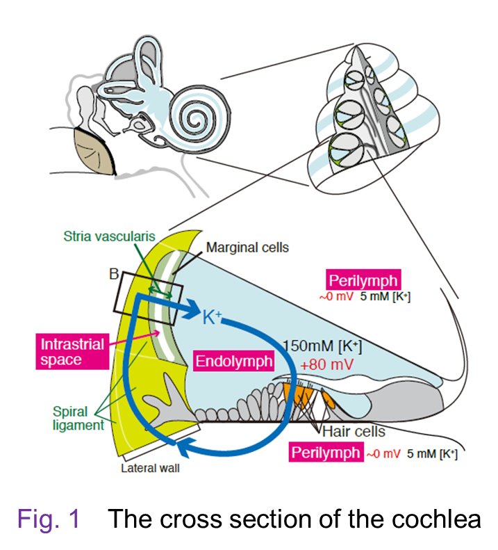 Figure 1 show a cross section of the cochlea, which is made up of three tubular
structures. Upper and lower tubules contain ‘perilymph’, which resembles
a regular extracellular solution and blood plasma. On the other hand, center
tubule is filled with an unusual extracellular solution, ‘endolymph’, which
shows 150 mM [K+] and 2 – 5 mM [Na+]. This profile looks like the properties in an intracellular solution. In addition, the endolymph always exhibits a highly positive potential of +80 – +100 mV with reference to the perilymph. These unique electrochemical properties are essential for high sensitivity observed in mammalian hearing and their disruption results in deafness. Take a look at
the other page to learn how the cochlea works.
Figure 1 show a cross section of the cochlea, which is made up of three tubular
structures. Upper and lower tubules contain ‘perilymph’, which resembles
a regular extracellular solution and blood plasma. On the other hand, center
tubule is filled with an unusual extracellular solution, ‘endolymph’, which
shows 150 mM [K+] and 2 – 5 mM [Na+]. This profile looks like the properties in an intracellular solution. In addition, the endolymph always exhibits a highly positive potential of +80 – +100 mV with reference to the perilymph. These unique electrochemical properties are essential for high sensitivity observed in mammalian hearing and their disruption results in deafness. Take a look at
the other page to learn how the cochlea works.
The milieu of the endolymph has been considered to be maintained by K+-circulation or K+-recycling between this fluid and the perilymph. This unidirectional ion transport is likely to be driven by ‘lateral cochlear wall’, an epithelial-like tissue composed of inner and outer layers. However, it has remained uncertain how the K+-circulation actually contributes to the endolymph in vivo. We have focused on the high potential of the endolymph and analyzed the mechanism underlying formation of this electrical element for a long time. Together with the results reported by other groups, our research has revealed the processes described in the followings. Firstly, the K+-circulation depends on functional coupling of ion channels and transporters
in the lateral wall (Fig. 2). 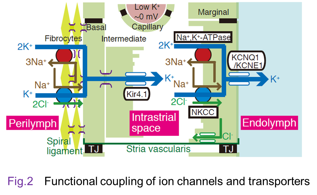 Secondly, the origin of the endolymphatic potential is lateral-wall’s
two different concentration cells that are connected in series and these
batteries are controlled by the K+-circulation. Thirdly, these two issues are integrated in vivo, resulting
in maintenance of the endolymphatic potential [4-12]. Moreover, interference
with a crucial factor involved in the K+-circulation by optogenetic approach caused repetitive acute hearing loss
observed in clinical sites [3].
Secondly, the origin of the endolymphatic potential is lateral-wall’s
two different concentration cells that are connected in series and these
batteries are controlled by the K+-circulation. Thirdly, these two issues are integrated in vivo, resulting
in maintenance of the endolymphatic potential [4-12]. Moreover, interference
with a crucial factor involved in the K+-circulation by optogenetic approach caused repetitive acute hearing loss
observed in clinical sites [3].
By collaborating with engineers, we will develop advanced methodologies to clarify the mechanisms underlying the electrochemical and other profiles of the endolymph and their physiological and pathological significances.
【Experimental procedures】
Go to the page entitled ‘Analytical instruments’ to see the details.
(1) Molecular biological and histological methods[3, 4, 12]:These methods are used to identify the molecular basis and tissue and
cellular localization of the ion channels and transporters involved in
driving the K+-circulation.
(2) Comprehensive analysis of the proteins[13]:With comprehensive approaches using HPLC and LC-MS/MS, we are working
on clarification of the profile of the proteins that contribute to the
K+-circulation. This project is carried out by collaborating with Prof. Shushi Nagamori in Nara.
(3) Ion selective microelectrodes [4-10]:The K+-circulation maintains the high potential in the endolymph by regulating [K+] in the lateral wall. Therefore, it is crucial for the project to examine
the dynamics of K+ profile. We fabricate by ourselves ion selective microelectrode that monitor
the potential and [K+] in the microenvironment ‘simultaneously and in real time’ and use this sensor for in vivo measurements.
(4) Computer simulation [4, 9]:Some of vital phenomena cannot be measured by any experimental
techniques. Indeed, in this project, we are unable to directly detect ionic
flows through the channels or transporters in vivo. This issue is accessible to theoretical approaches. We have constructed
a number of equations that represent the ion channels and transporters
in the lateral wall and integrated these ionic flows to those in sensory
hair cells in silico. This ‘Nin-Hibino-Kurachi (NHK) model’ can reconstitute the K+-circulation as well as the electrochemical properties of the lateral wall
and endolymph in different conditions [9]. This model has recently renewed
with the experimental observations (fi-NHK model) (for source code, see the linkpage) [4]. Our model can show the processes
that might be critically involved in the vital events but invisible in
the experimental approaches. We will further improve the model via feedback
from the experimental results and use for a variety of projects in the
future.