

Analytical instruments
We construct or develop majority of key analytical apparatus by ourselves and carry out unique measurements. We also perform a variety of molecular biological and histological experiments.
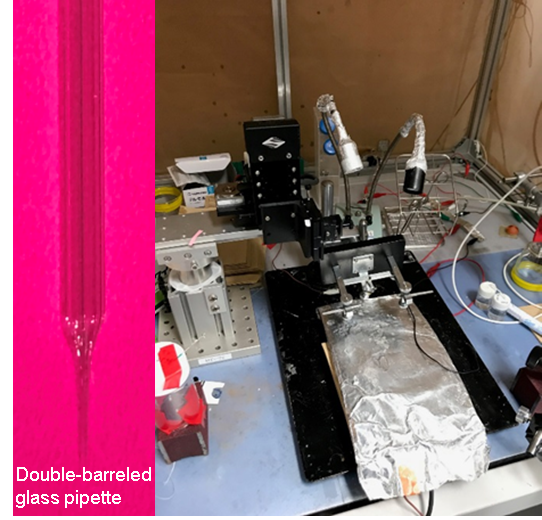
(A) Ion selective electrodes→Analysis of ‘endolymph’
Ion selective microelectrodes are fabricated in our lab from commercially available double-barreled glass capillaries. Tip diameter of the microelectrode is 1 – 3 µm. The tip of one barrel is filled with a hydrophobic, K+-selective material. The other barrel contains an electrolyte solution. The former measures [K+], whereas the latter monitors potential. In other words, this microelectrode permits us to detect changes of ion concentrations and potential in the microenvironment ‘simultaneously and in real time’. To our knowledge, we are currently only one group that can examine the electrochemical properties in the cochlea with the double-barreled ion selective microelectrode while perfusing drugs to the inner ear.
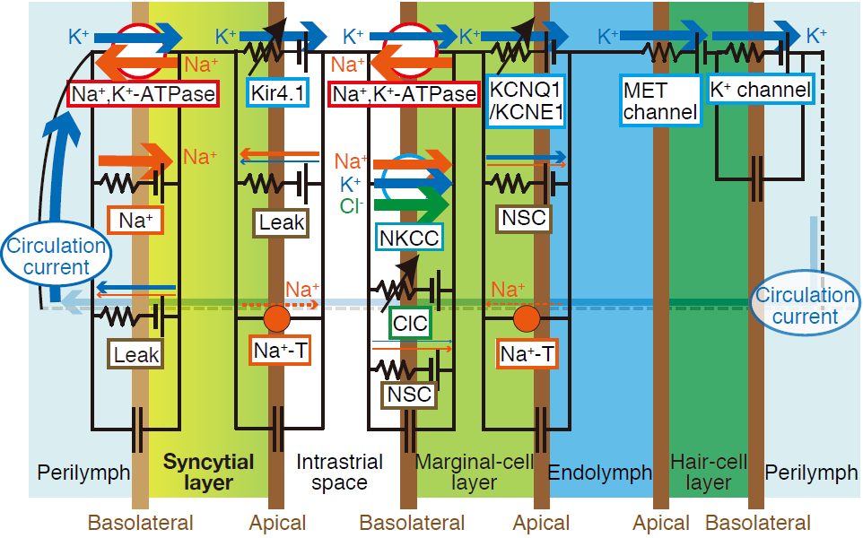
(B) Computer simulation
On the basis of the electrophysiological and histological data obtained by ourselves as well as other groups, we have developed a computer model that reconstitutes the K+-circulation between the perilymph and endolymph. In details, the model is an electrical circuit made up of different equations that show activities and functions of the ion channels and transporters contributing to the K+-circulation. The circuit also represents the number of cells and their morphology in a cross section of the cochlea. This called ‘Nin-Hibino-Kurachi (NHK) model’ can simulate the potential and ion concentrations in the extracellular and intracellular compartments not only under normal conditions but also when the animal’s hearing is disrupted by drugs or diseases.
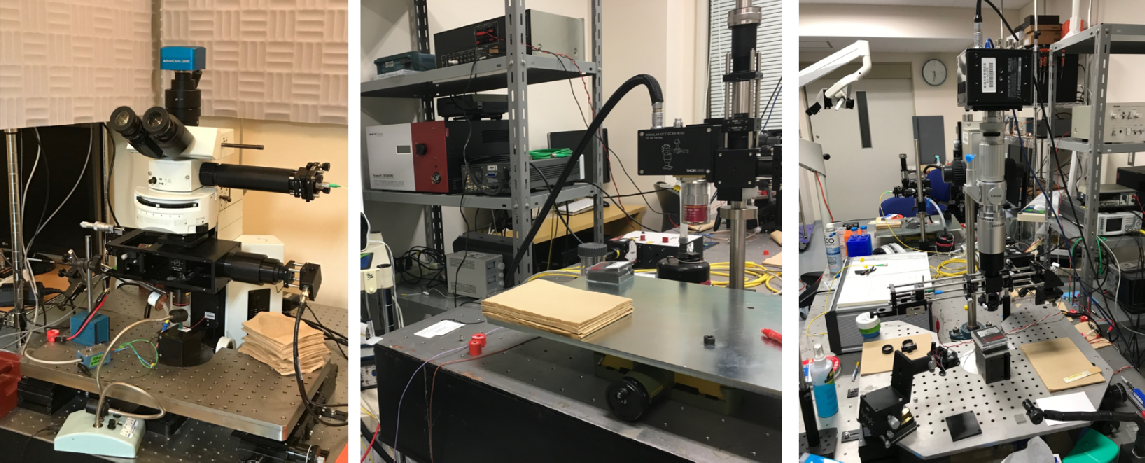
(C) Vibrometries →Nanometer-scale motions in the cochlear sensory epithelium
We have a couple of different vibrometries to measure nanometer-scale motions in the cochlear sensory epithelium. The setups and computer programs have been made by ourselves.
The laser interferometer described in the left panel can measure the sample’s vibration at ~40
pm. However, this system detects the motion at one point of the sample
and it is unable to take any tomographic images. The methodology and algorithm
for the measurements in our system are different from those in commercially
available interferometers so that it can detect a particular parameter.
MS en-face optical coherence tomography (OCT) shown in the right panel allows us to take 3D image of the sample without
any fluorescent labeling and detect at once the nanometer-scale vibrations
in a planner full-field. This state-of-the-art apparatus is being developed
by Dr. Samuel Choi, an assistant professor in Department of Engineering,
Niigata University. We are currently making every effort to improve the
performance of the en-face OCT.
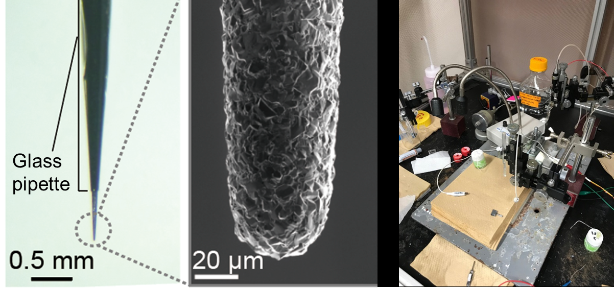
(D) An advanced drug monitoring system→ Advanced drug monitoring system
The drug monitoring system developed by our group is equipped with two different microsensors: a needle-type diamond microsensor (tip diameter; ~40 µm) fabricated by Einaga’s group in Keio University and a classical glass microelectrode (tip diameter; ~1 µm). This arrangement permits us to detect the local pharmacokinetic and pharmacodynamics ‘simultaneously and in real time’.
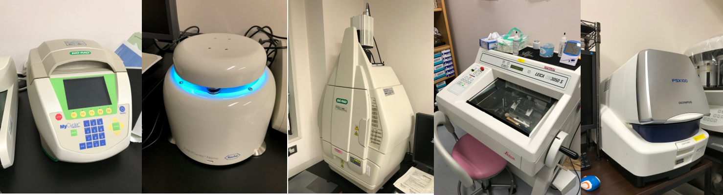
(E)Molecular biological and histological instruments
We have instruments for PCR, real-time PCR, and western-blot (chemidoc), a cryostat, and an all-in-one type fluorescent microscope. These machines were purchased from commercial lines. Our lab also harbors a cell culture room.