

Mechanism underlying transmission of acoustic signals in the inner ear
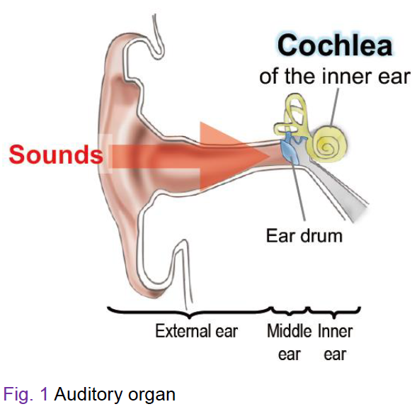 Sounds from the outside are propagated through the outer and middle ears
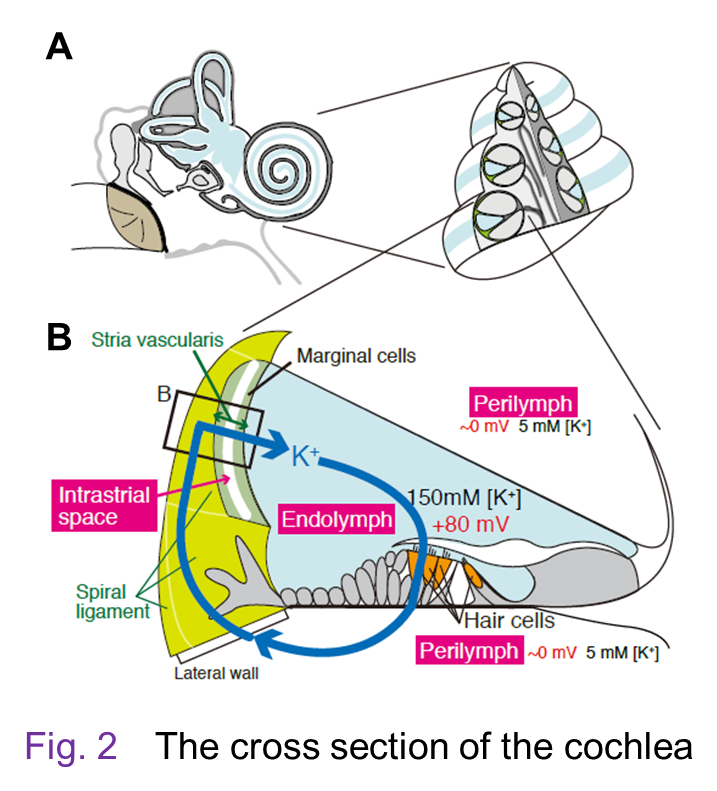
and reach the cochlea of the inner ear (Fig. 1). Figure 2A illustrates the cross section of the cochlea. This organ has three tubular
structures. Upper and lower tubules contain a perilymph, whose ion composition
is similar to the regular extracellular solution such as blood plasma (i.e.,
150 mM [Na+] and 5 mM [K+]). On the other hand, the center tubule is filled with a unique extracellular solution, ‘endolymph’, which shows high [K+] of 150 mM, low [Na+] of 2 – 5 mM, and low [Ca2+] of 20 µM. Moreover, the endolymph exhibits a highly positive potential
of +80 – +100 mV with reference to the perilymph. These electrochemical
properties are observed only in the cochlea but not detected in any other
organs.
Sounds from the outside are propagated through the outer and middle ears
and reach the cochlea of the inner ear (Fig. 1). Figure 2A illustrates the cross section of the cochlea. This organ has three tubular
structures. Upper and lower tubules contain a perilymph, whose ion composition
is similar to the regular extracellular solution such as blood plasma (i.e.,
150 mM [Na+] and 5 mM [K+]). On the other hand, the center tubule is filled with a unique extracellular solution, ‘endolymph’, which shows high [K+] of 150 mM, low [Na+] of 2 – 5 mM, and low [Ca2+] of 20 µM. Moreover, the endolymph exhibits a highly positive potential
of +80 – +100 mV with reference to the perilymph. These electrochemical
properties are observed only in the cochlea but not detected in any other
organs.
 In the cochlea, sensory hair cells expose their apical membrane, which
has the stereocilia, to the endolymph and bathe their cell bodies in the
perilymph. The hair cells as well as supporting cells beneath them lie
on the ‘basilar membrane’, an extracellular matrix. A unit composed of
these three elements is called ‘sensory epithelium’. Acoustic inputs firstly
stimulate the basilar membrane and vibrates the sensory epithelium. This
event deflects the stereocilia of the hair cells and further activates
‘mechanoelectrical transduction channels’ at their tips. The channel’s
opening enters K+ in the endolymph into the hair cells and electrically
excites them. In this process, mechanical energy of sounds is transduced to electrical signals. Finally, neurotransmission occurs in the cell bodies of hair cells and
acoustic information is conveyed to the brain through neural fibers.
In the cochlea, sensory hair cells expose their apical membrane, which
has the stereocilia, to the endolymph and bathe their cell bodies in the
perilymph. The hair cells as well as supporting cells beneath them lie
on the ‘basilar membrane’, an extracellular matrix. A unit composed of
these three elements is called ‘sensory epithelium’. Acoustic inputs firstly
stimulate the basilar membrane and vibrates the sensory epithelium. This
event deflects the stereocilia of the hair cells and further activates
‘mechanoelectrical transduction channels’ at their tips. The channel’s
opening enters K+ in the endolymph into the hair cells and electrically
excites them. In this process, mechanical energy of sounds is transduced to electrical signals. Finally, neurotransmission occurs in the cell bodies of hair cells and
acoustic information is conveyed to the brain through neural fibers.
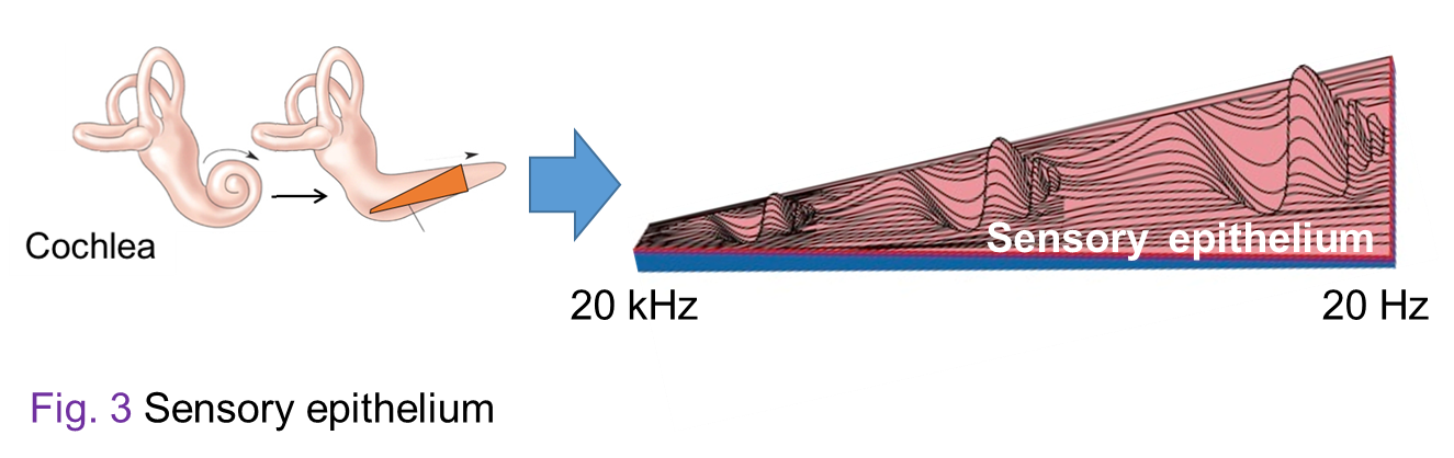
Young people who have normal hearing can perceive sounds that range from 20 Hz to 20,000 Hz (i.e., 20 kHz). This ‘tonotopic organization’ stems from multiple elements; here let us show one of them. Displayed in Figure 3 is the inside of an uncoiled cochlea. In this organ, apex portion is sensitive to low pitch sounds, whereas base portion responds to high pitch sounds. This characteristics depends on property of the basilar membrane. The membrane is getting softer and thinner as you are approaching to the apex portion. This physical gradient plays key roles in tonotopic organization. The tonotopy also involves other machineries.