Current Reserch
1. The principle of voltage sensor
For a long time, studies on the mechanisms of neuron excitation and impulse transmission have been the major theme in physiology. In the mid of 20th century Huxley and Hodgkin discovered that sodium ions play an essential role in the formation of action potential in the giant axon of the squid. They quantitatively measured the ion currents and showed that,
(1) Na+ and K+ go through different pathways in plasma membrane.
(2) The opening and closing of these pathways are quantitatively described by operation of voltage-gated ion channels which are time and membrane potential dependent and action potentials were numerically reconstituted based on these elements.
Ion channels, including voltage-gated sodium channels and potassium channels, were characterized in detail by using toxins and measuring microscopic currents by patch clamp technique. In late 20th century, molecular cloning techniques led to identification of these ion channel molecules. In the 21st century the structure of voltage-gated ion channels at atomic level was resolved by X-ray crystallography. This led to clear understanding of the channel molecules.
In the years 2005 and 2006, we discovered two unique voltage dependent membrane proteins (Murata et al, Nature, 2005; Sasaki et al, Science,2006). One of the proteins, identified from the ascidian (Ciona intestinalis) was named Ci- voltage sensing phosphatase (Ci-VSP). This protein consists of two modules; the voltage sensor and phosphatase. Upon depolarization the voltage sensor undergoes conformational change which is coupled to the enzymatic activity of the phosphatase region. This enzyme dephosphorylates phosphatidyl inositols which are involved in various signalling functions within the cell. Therefore electrical signals are converted to chemical signals by VSP.
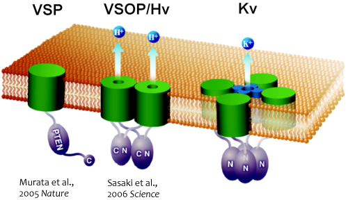
Fig.1 The new types of voltage sensor proteins
VSP : Voltage sensor phosphotase, VSOP : Voltage-gated proton channel, Kv : Voltage-gated potacium ion channel
The other protein was named VSOP/Hv1 (VSOP=voltage sensor only protein). This protein consists of only the voltage sensor domain and lacks an authentic pore domain. Although it lacks the pore, it shows proton-selective voltage-dependent conductance .The voltage sensor domain of VSOP has a double role in playing i.e voltage-dependent gating and proton permeation. Both "gating" and "ion permation" are two central questions in the field of ion channels. In this sense, VSOP/Hv1 provides a good model to understand general principle of operation of ion channel.
The Hv channel consists of two monomers which associate through cytoplasmic coil-coil interaction (Koch et al, PNAS, 2008, in collaboration with Dr Larsson of Miami University). X-ray crystallographic analysis of the cytoplasmic region of VSOP showed that the mechanism of opening and closing is precisely controlled by the coil-coil structure interactions which occur at normal body temperature (Fujiwara et al, Nat. Commun, 2012).
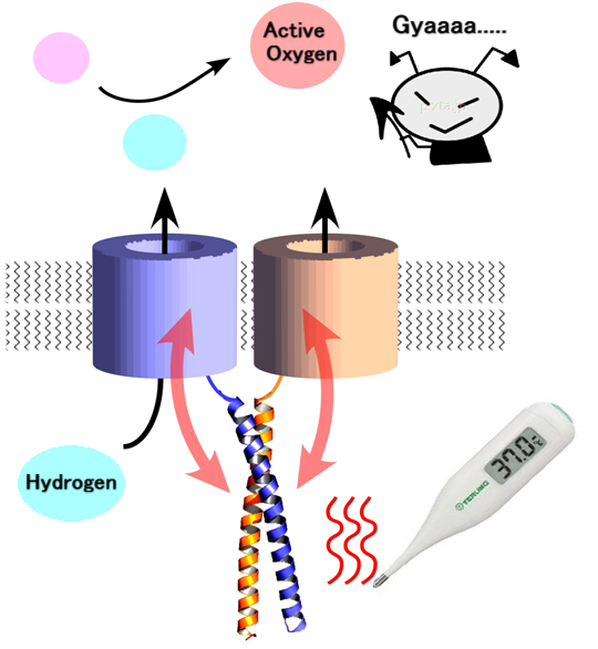
Fig.2 The role of coiled coil in voltage-gated proton channel
1. How can VSP phosphatase activity be activated by the operation of voltage sensor?
2. How can change in membrane potential make VSOP/Hv1 proton conductive?
To address these questions, molecular mechanisms have been intensively investigated by combining electrophysiological and optical techniques.
2. Molecular mechanisms and structural biology of phosphoinositide phosphatases
Many studies have been done on the molecular mechanisms of cancer and metabolic abnormalities. Tyrosine phosphatases and PTEN have been shown to play a role in development of these diseases, VSP has the cytoplasmic enzyme region showing similar structure to these enzymes, and its enzymatic activity is controlled by changes in membrane potential. Therefore elucidating the mechanisms by which this change takes place, it will lead to understanding the mechanisms of tyrosine phosphatases and PTEN in cancer and its related diseases.
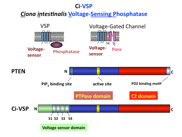
Fig.3 VSP and PTEN
Enzymatic domain of VSP has a high similarity with PTEN and both enzymes target phosphoinositides as substrates (Iwasaki et al.,PNAS, 2008; Matsuda et al, JBC, 2011). In addition, it was shown that VSP has a unique characteristic whereby the substrate specificity alters with changing membrane potential which is not a common feature of many enzymes (Kurokawa et al, PNAS, 2012).
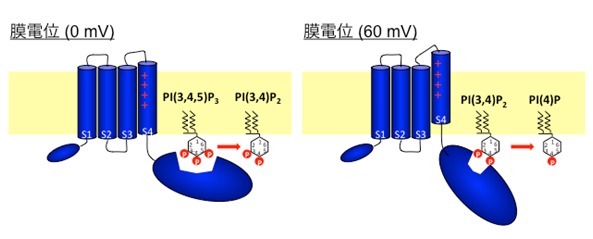
Fig.4 VSP function
3. Role of voltage-gated proton channels in blood cells and microglia in brain
VSOP/Hv1 is conserved from marine invertebrates to human. In mammals these channels are expressed in blood cells such as macrophages and neutrophils and microglia. In many cells including osteoclasts, ATP hydrolysis by proton pump (V-ATPase) results in production of protons which are transported to the extracellular side. It is well known that Hv channels are able to export large quantities of protons from the cell more accurately than proton-pump pathways. In most cases Hv can only be activated when the membrane potential is more positive than the reversal potential for proton flux. This is due to intrinsic properties of pH-regulated gating of Hv channels. Vectoral transport of proton to extracellular side can be achieved without requiring the use of ATP.
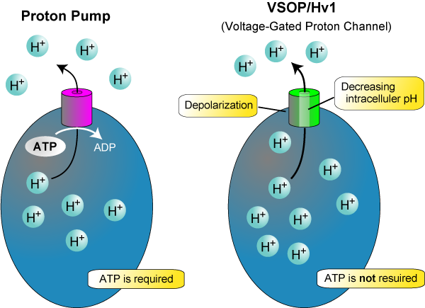
Fig.5 The role of voltage-gated proton channnel
Until now, using electrophysiological studies, it has been suggested that Hv channel promotes production of reactive oxygen species in phagocytic cells. Initially, superoxide anions, O2- are produced by NADPH oxidase, which is a protein complex consisting of the membrane protein, gp 91 and other auxilary subunits. O2- is secreted into the lumen of phagosome or extracellular side. If this situation continues, the cell membrane potential is increased due to imbalance of the charge, resulting in inhibition of the oxidase and attenuated production of O2-. This charge imbalance is corrected by the proton transport through Hv channel. We succeeded in identifying molecular properties of this channel (Sasaki et al, Science, 2006). In our analysis of knockout mouse VSOP/Hv, it was found that Hv channel controls the pH of cytoplasm and membrane potential due to proton transport leading to continued production of reactive oxygen species (Okochi et al, BBRC, 2009, El Chemaly et al, JEM, 2010).
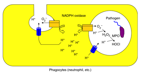
Fig.6 The role of voltage-gated proton channel on phagocyte
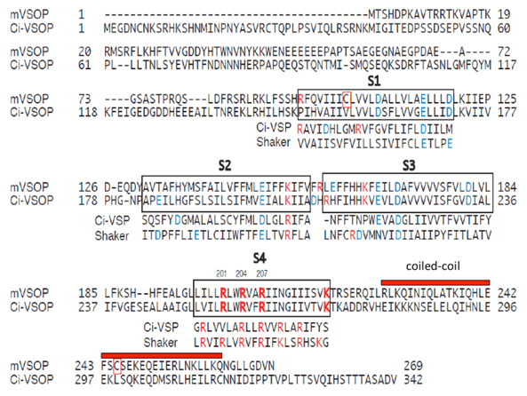
Fig.7 The comparison of sequence of voltage-gated proton channel
4. Molecular tool development for the visualization of membrane potential.
Neuronal circuit is the key mechanisms of brain function. To understand operation mechanisms of neuronal circuits, it is very important to simultaneously measure the activity of individual neurons. Further, electrophysiological approaches by use of glass electrodes to cells and tissue cultures are relatively tedious. It will be useful to visualize the membrane potential and behaviour of multiple neurons without using invasive method. So far, potential sensitive organic dyes have been used as voltage probe. However these probes are difficult to be targeted to a specific cells and another drawback is that signal strength is weak. Recently, several probes have been made by linking the voltage sensor of VSP to fluorescent proteins. One such probe has been developed by assistant professor Dr Tsutsui who by then was enrolled in the research group of Dr. Miyawaki of RIKEN. The probe was named Mermaid (a combination of proteins derived from marine organisms). The probe exhibited large changes in fluorescence in the imaging of cultured cells such as single neuron and cardiac cells (Tsutsui et al., Nature Methods, 2008). Presently, we are trying to improve the membrane potential molecular probe technology toward the future versatile in vivo measurement of electrical activities.
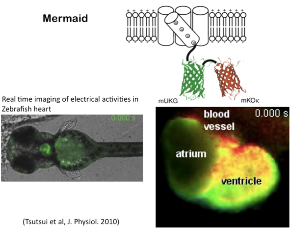
Fig.8 The development for visualizing membrane potential
5. Elucidating new roles of membrane potential signals in biological phenomena
Studies on voltage sensor protein have led to the discovery of new molecular pathways that accompany changes in membrane potential. This has necessitated the need to review the physiological role of membrane potential. For example, polyspermy block at fertilization membrane potential, involvement of changes in membrane potential in the left-right nodal determination and regeneration in non-mammalian vertebrates have been suggested despite the fact that clear molecular mechanisms are not well understood. Currently, with other collaborators we are working hard to find out the physiological role of VSP and other voltage sensitive properties. Moreover, use of voltage-based probes will facilitate elucidation of unaddressed roles of changes in membrane potential.
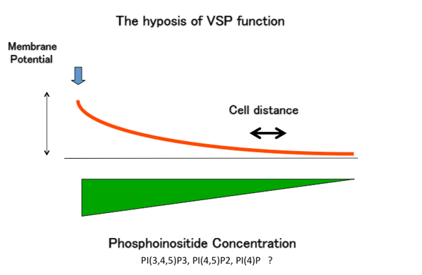
Fig.9 VSP function
In VSP, the enzymatic activity varies with the changing membrane potential. So far its physiological role is not well understood. Since VSP is expressed in testis, it may possibly play an important role in the development of sperm. The figure above points potential physiological role of VSP based on its molecular properties. If the VSP is present in cell membrane uniformly, then graded concentration of phosphoinositols will take place depending on the gradient of the potential.
6. Fundamental studies of visual function recovery based on artificial retina. (Dr. Tomomitsu Miyoshi)
In diseases such as retinitis pigmentosa, the cells are modified. By electrically stimulating the unaffected nerve cells, it is possible to restore perception of light. However, a limited number of electrodes denature retina during electrical stimulation, using different methods some of the challenges have been overcomed. In addition we have analyzed the physiological effects and found that electrical stimulation is neuroprotective. Research and development of artificial retina is being undertaken by experts from different fields of study such as medicine, space engineering, ophthalmology.