|
|
|
|
HOME > Proposed research projects > 2012-2013: Proposed research projects 13
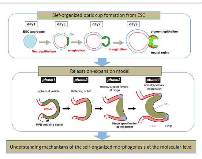
 2012-2013: Proposed research project 13Molecular mechanisms involved in the self-organized optic cup formation in vitro
Purpose of the Research ProjectIn the field of embryology, how complex vertebrate organs are determined and shaped is a matter of keen interest. Our understanding of the dynamic processes of organogenesis is still limited and often fragmental. Recent progress in ESC culture studies has enabled the use of this in vitro system for analyzing various steps of mammalian neural tissue development. We have recently reported that the optic cup structure is generated by self-organization in 3D culture of mouse ESCs. Based on the self-organizing phenomenon, we proposed the "relaxation-expansion" model to mechanically interpret the tissue dynamics that enable the spontaneous optic cup formation. This model involves only three local rules (relaxation, apical constriction and expansion), and its computer simulation recapitulates the optic-cup morphogenesis in silico. To understand the domain- and phase-specific organization of the local rules, it will be important to combine the tissue-level models with some underlying molecular-level models, in particular for, cytoskelton regulations. In this project, I aim to clarify cytoskelton dynamics and its regulation during the self-organized optic cup morphogenesis in vitro. Content of the Research ProjectTo clarify the mechanism underlying the self-organized optic cup morphogenesis at the cellular and molecular level, I will focus on the following two aspects. 1, 4D-imaging study for understanding cytoskelton dynamics during the self-organized optic cup formation. 2, How actomyosin activity is spatiotemporally controlled during the optic cup formation? Expected Research Achievements and Scientific SignificanceProgress in understanding the mechanism of the self-organized optic cup formation at the cellular and the molecular level would contribute to comprehend general mechanisms of epithelial deformation, and to develop a novel methodology for the tissue engineering. In addition, functional epithelial tissues with proper 3D shape generated from human ESC/iPS cells are attractive resources not only for regenerative medicine, but also for various biomedical applications including drug discovery, toxicology and pathogenesis studies. Further advancement in understanding mechanisms involving local intercellular interactions would open avenues for a wide variety of stem cell applications.
|