


わたちたちは、独自に開発や最適化を進めている特殊計測系をはじめ、多彩なアプローチを用いて研究しています。
ナノ振動計測装置研究:感覚上皮帯のナノ振動
左側のレーザー干渉計は、断層撮影はできませんが、振動を20ピコメートルまで高精度に測定することができます(非生物を使った実験)。ただ、1点のみの情報取得に限ります。この装置の測定方法は、一般的な市販の干渉計とは異なっており、特殊な動的パラメータが捉えられます。装置やプログラムは、全て自作です。
中央のOptical Coherence Tomograpy(OCT)は、いわば「光エコー」です。ソーラボ社の機器を大改造しています。一点のレーザ照射によって、z軸方向(深部方向)の断層撮影が可能です。このレーザをx方向やy方向へスキャンすることによって3次元的な画像を取得します。精密な振動計測を目指し、日々、改良しています。
右側のen-face OCTは、もう一つの「光エコー」です。xy方向に平面一括撮像しながら、振動を計測できます。z軸方向に断層も可能です。新潟大学工学部の崔 森悦が中心になって開発しました。現在、より性能を上げる努力をしています。
また、OCTに関しては、より画像を鮮明にするため、現在、補償光学の技術を導入しつつあります。

イオン電極法研究:生体電池の仕組み
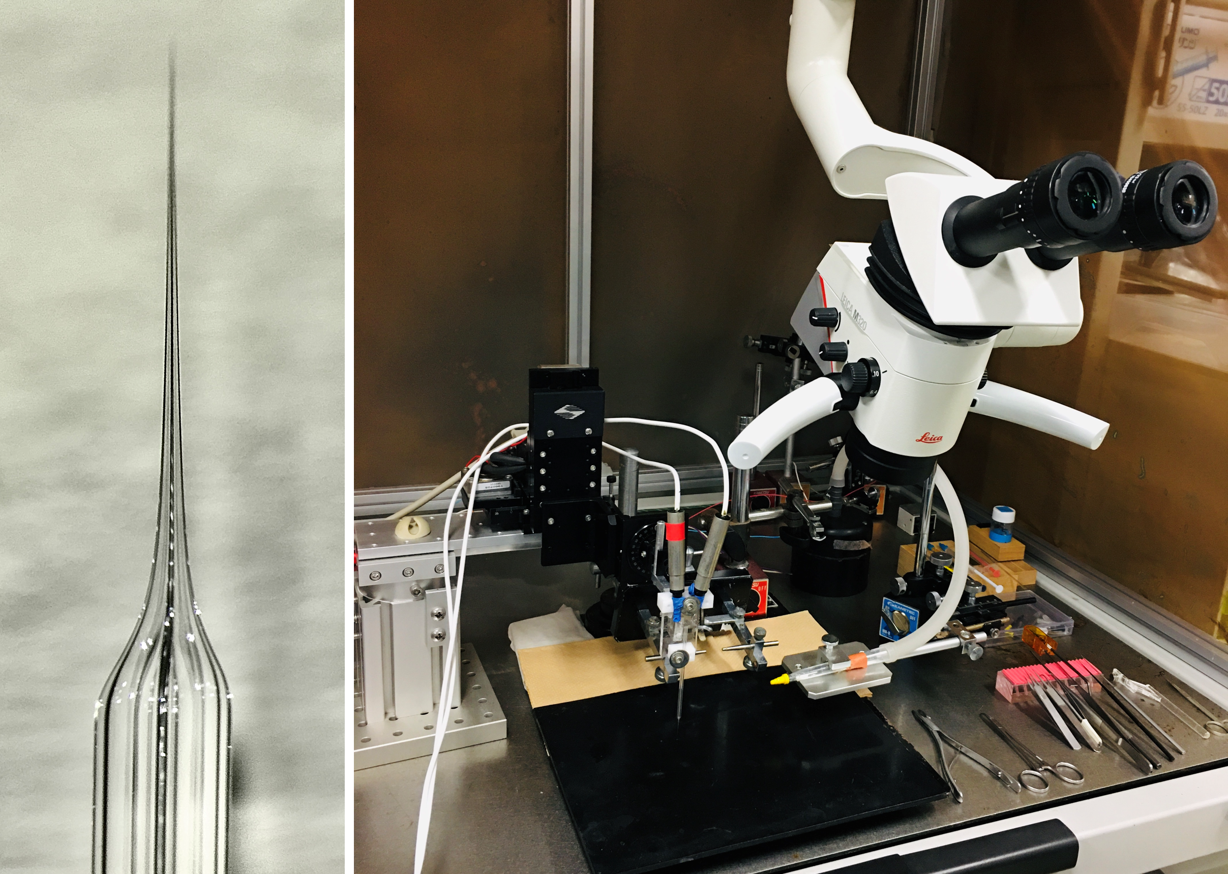
ガラス2連管を細く引いて電極を作ります(図の左側)。先端は1~3マイクロメートルです。一方の管の先端にK+を選択的に通す特殊な疎水性の物質を詰めます。もう一方の管には、通常の電解質溶液を入れます。前者でK+濃度、後者で電位を計測します。つまり、“その場”の濃度と電位が同時にリアルタイム測定できます。内耳蝸牛に様々な薬物を灌流しながらイオン電極を遂行できるのは、現在、世界でわたしたちのみです。図の右のように、手術用顕微鏡を使いながら、電極を内耳蝸牛にアプローチしていきます。音刺激しながら測定することもできます。
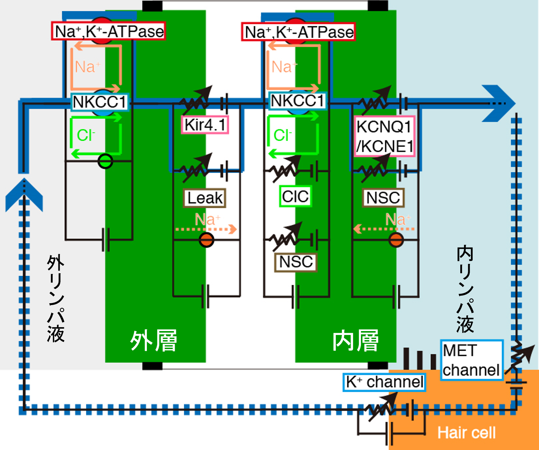
コンピューターシュミレーション研究:生体電池の仕組み
わたしたちや他のグループの実験結果に基づき、内耳蝸牛の生体電池やイオン・電位環境を再現するコンピュータモデルを構築しました。ここでは、イオンを輸送するチャネルやトランスポータの働きを数式で表し、それらをつなぐことでバーチャルな電気回路を作成しています。この「Nin-Hibino-Kurachi(NHK)モデル」を用いると、健康な聴力状態のみならず、音を聴いた場合や、薬物や病気で難聴になっている際の蝸牛の細胞内外のイオン・電位環境がシミュレーションできます。
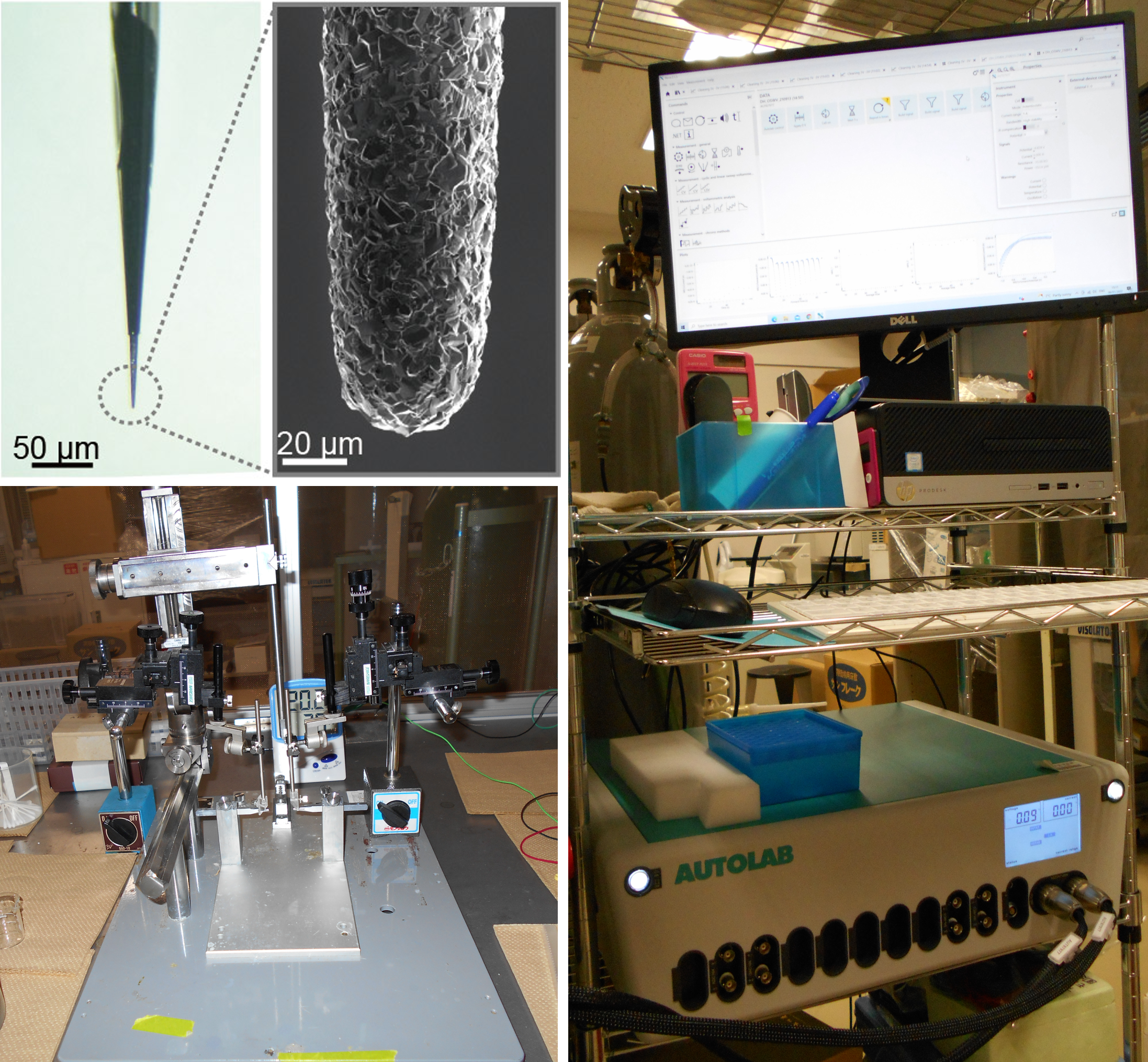
薬物モニタリングシステム薬物モニタリングシステム
わたしたちが構築した薬物モニタリングシステムは、慶應大学理工学部の栄長グループが創製する「針状ダイヤモンド電極センサ」(先端径40マイクロメートル:図の左上)と古典的な「微小ガラス電極センサ」(先端径1マイクロメートル)からなります。ダイヤモンドセンサで薬物の濃度の変化を、ガラス電極センサで細胞や組織の電気活動を追尾します。これら2つのパラメータ、すなわち、Pharmacokinetics(PK)とPharmacodynamics(PD)を同時にリアルタイムで捉えることができる、世界唯一のシステムです。現在、針状ダイヤモンド電極センサをさらに微細化するなど、改良を進めています。図の左下は生体計測用のステージ、右はポテンシオスタットと呼ばれる特殊なアンプです。
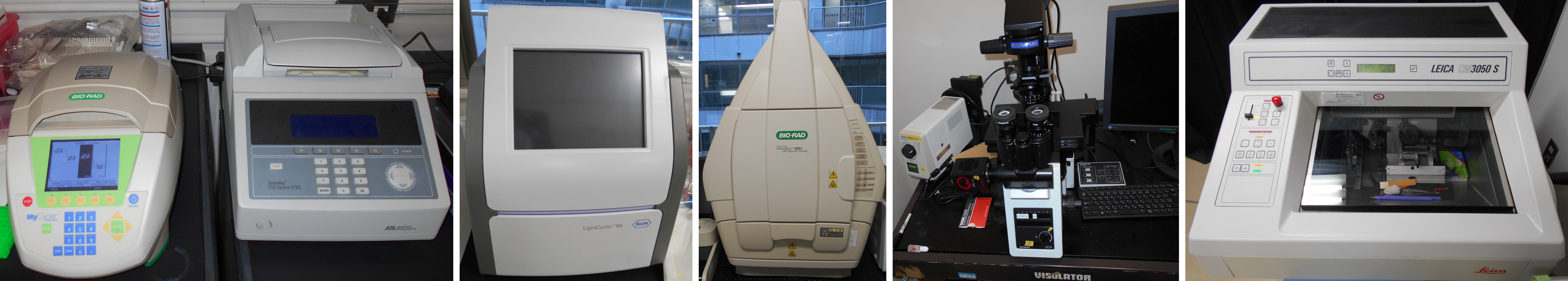
分子生物学的・生化学的・組織学的手法
PCR(2種類)、Real-time PCR、ケミドック、蛍光顕微鏡、クライオスタット、パッチクランプシステム(写真なし)、です。全て市販品です。他にも様々な機器があります。細胞培養室もあります。