


Cochlea and Hearing Loss
❶ Nanoscale Vibrations in Cochlear Sensory Epithelium
【Outline of the Research Project】
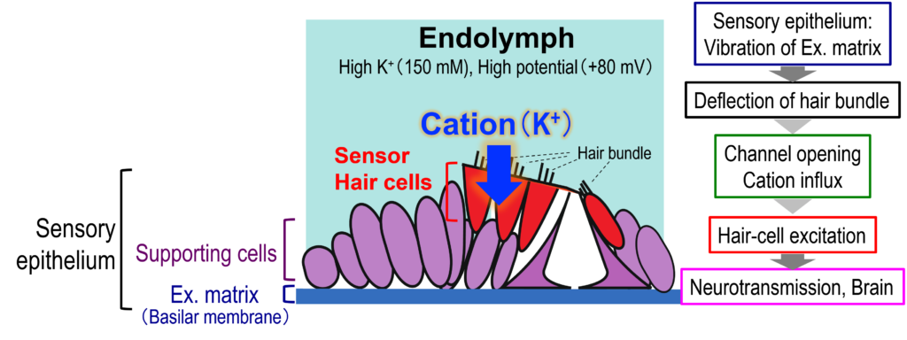
The cochlea of the inner ear has the sensory epithelium, an essential tissue made up of three layers, i.e., hair-cell layer, supporting-cell layer, and extracellular matrix layer called the ‘basilar membrane’ (Fig. 1). Acoustic inputs firstly stimulate the basilar membrane and vibrates the sensory epithelium. This event deflects the stereocilia of the hair cells and further activates ‘mechanoelectrical transduction channels’ at their tips. The channel’s opening enters K+ in the endolymph into the hair cells and electrically excites them. In this process, mechanical energy of sounds is transduced to electrical signals. For the details of the process, see the section ‘How Does the Cochlea of the Inner Ear Work?’
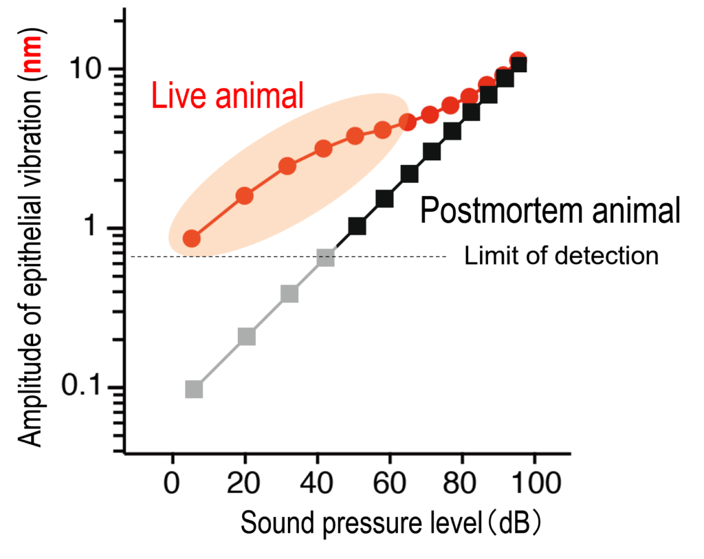
Our hearing can enhance small sounds and perceive them with higher sensitivity, whereas it detects loud sounds with lower sensitivity. This property emerges primarily from a unique pattern of the motion in the sensory epithelium. Figure 2 plots the amplitude of epithelium’s vibration as a function of intensity of the acoustic stimuli. The relationship described in this panel clearly represents the aforementioned property. This is referred to as ‘nonlinear response’ or ‘nonlinear amplification’, which disappears when the animal is dead. Note that the amplitude of the vibration in the epithelium is very subtle and ranges from 0.1 nm at minimum to 10 nm at maximum (Fig. 2); the difference is 100-fold. Your hearing can sense a buzzing of a mosquito that flies 3 m away from you as well as accept a jet engine sound emitted form an aircraft nearby. This difference of 1,000,000-fold in sound pressure levels is highly compressed to 100-fold in the motion in the sensory epithelium. The large dynamic range results from the nonlinear response mentioned above and this profile is crucially involved in establishment of the sharp tuning of our hearing.
Sensory hair cells are classified into two groups, outer and inner hair cells. Outer hair cells isolated from the cochlea can change their shape in response to electrical stimuli. This motility is likely to crucially contribute to the nonlinear response in the sensory epithelium. Nevertheless, it has not yet been fully elucidated ‘in vivo’ how the hair cells move and their motion are associated with vibrations in the supporting-cell layer and/or basilar membrane. To address these issues, we are developing an advanced laser interferometer and a unique optical coherence tomography (OCT) together with engineers and investigating the mechanisms underlying the characteristic vibrations in the sensory epithelium. For details, see the page of ‘Analytical Instruments’. We have also begun to study the pathological relevance of disorder of the vibrations to hearing loss.
Publication List
(1) Yui H, Fujiyama Y, Urashima S, Ota T, Hibino H (2026). Off-axis illumination and epi-collection confocal Brillouin scattering micro-spectroscopy. Anal Chem, in press.
(2) Nin F*#, Choi S*, Ota T, Qi Z, Hibino H (2021). Optimization of spectral-domain optical coherence tomography with a supercontinuum source for in-vivo motion detection of low reflective outer hair cells in guinea pig cochleae. Optical Review 28, 239–254. [*: equal contributors, #: corresponding author]
(3) Ota T#, Nin F#*, Choi S, Muramatsu S, Sawamura S, Ogata G, Sato MP, Doi Kat., Doi Ken., Tsuji T, Kawano S, Reichenbach T, Hibino H (2020). Characterisation of the static offset in the travelling wave in the cochlear basal turn. Pflügers Archiv - European Journal of Physiology 472, 625-635. [#: equal contributors][*: corresponding authors].
(4) Choi S, Nin F, Ota T, Sato K, Muramatsu S, Hibino H (2019). In vivo tomographic visualization of intracochlear vibration using a supercontinuum multifrequency-swept optical coherence microscope. Biomedical Optics Express 10(7), 3317-3342.
(5) Choi S, Sato K, Ota T, Nin F, Muramatsu S, Hibino H (2017). Multifrequency swept optical coherence tomographic vibrometry in biological tissues. Biomed Opt Express 8(2)608-621.
(6) Choi S#, Maruyama Y, Suzuki T, Nin F, Hibino H, Sasaki O (2015). Wide-field heterodyne interferometric vibrometry for two-dimensional surface vibration measurement. Opt Commun 356C:343-349.
(7) Choi S#, Watanabe T, Suzuki T, Nin F, Hibino H, Sasaki O (2015). Multifrequency swept common-path en-face OCT for wide-field measurement of interior surface vibrations in thick biological tissues. Opt Express 23(16):21078-21089.