Orthopedic Biomaterial Science
- Elucidation of the 3D jaw kinematics using CT and X-ray fluoroscopic images
- 3D dynamic analysis of limb joints, spine, and sacroiliac joints
- Elucidation of the 3D joint kinematics in patients with osteoarthritis of the knee wearing orthotic devices using X-ray fluoroscopic images and optical motion analysis system
- 3D dynamic analysis of limb joints, spine, and temporomandibular joints using MRI
- Development of a 3D human anatomy application
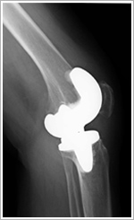
3D dynamic analysis of knee joints following total knee arthroplasty (TKA)
1)Post-arthroplasty dynamic images
Our originally developed analysis software enables multi-angle observation of knee joints after TKA.
This has allowed us for the first time to confirm if the surgery had been performed accurately and if the joints after surgery are moving properly. For artificial knee joints, it has been reported that post-operative knee flexion and durability (polyethylene wear) involve joint movement. Our study has revealed the detailed 3D movement of knee joints after TKA.
The 3D analysis has also helped unravel the mechanism of hip dislocation, which is a major issue after total hip arthroplasty, without examining actual cases, by providing accurate data on the bending degree and direction of the hip joint that affect dislocation. Moreover, the 3D analysis can be used for pre-operative joint evaluation for advance planning to achieve smooth post-operative joint movement.
2)Image creation principles
An X-ray imaging device (X-ray device in video mode) is used. Bone movement can be observed by moving the joint.
Using this device and applying the principle of shadow play, the original bone position can be calculated accurately.
 2D/3D registration technique
The accurate measurement of bone position is complex and impossible without using a computer technology. We have succeeded in achieving this measurement using our originally developed computer software.
3)Application in surgery
In the Orthopaedic Surgery of Osaka University, the 3D dynamic analysis has been available for pre-operative planning and post-operative evaluation, enabling physicians to perform surgery easier. In addition, patients can see the dynamics as a video to confirm how their joints move after surgery, which is highly valued.
The teamLabBody, a 3D human anatomy application, can be used widely by medical students, orthopedic surgeons, physical therapists, occupational therapists, Judo therapists, and many other medical specialists. For instance, it can be used to provide explanation to patients during medical treatment, obtain informed consent, and provide support during clinical practice and surgery.
We strongly hope that our originally developed software will contribute widely to medical care. We will provide cooperation as much as possible for the requests from patients such as detailed evaluation of joints before undergoing total arthroplasty and viewing of their post-operative joint movement as well as the requests from physicians for analysis of their surgical cases.