|
|
|
|
HOME > Proposed research projects > 2012-2013: Proposed research projects 21
 2012-2013: Proposed research project 21Tubulogenesis in the embryonic kidney by cytoskeletal regulation
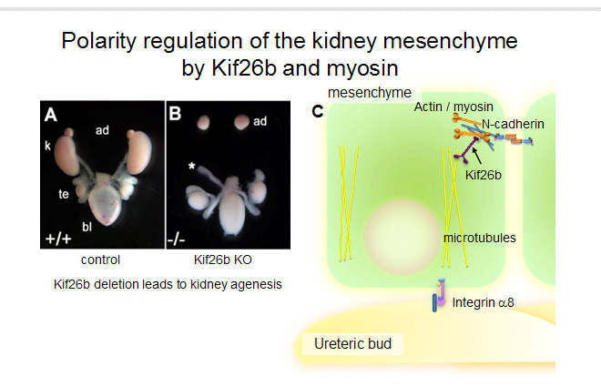
Purpose of the Research ProjectThe kidney is generated through mutual interactions between two precursor tissues: metanephric mesenchyme and ureteric bud. The mesenchyme is transformed to epilethelia and forms a lumen (epithelial-mesenchymal transformation, MET), and connected to the lumen derived from the ureteric bud to form a functional unit, the nephron. We identified a trasncrition factor Sall1 and its downstream target Kif26b both of which are expressed in the mesenchyme, and found that mice deficient for these genes lacked the kidneys completely. We further showed that microtubule-associated Kif26b bound the non muscle myosin heavy chain II (NMHC II, Myh9/10) and controled the polarity and adhesion of mesenchymal cells. The purpose of this study is to analyze the roles of cytoskeleton in the tubulogenesis, including MET, during kidney development. Content of the Research ProjectAs the conventional mutants of Kif26b and Myh9/10 exhibited too severe phenotypes and lethality, we plan to analyze the conditional knockouts of these genes to analyze the tubulogenesis in the embryonic kidney. We will generate the mesenchyme-specific mutants and examine the polarity of the mesenchyme and the MET process. We will utilize not only the standard histological approaches but also real-time fluorescent imaging techniques applied to the kidney organ culture. As Myh9/10 is also expressed in the ureteric bud where Kif26b is undetectable, we will also generate the ureteric-bud specific Myh9/10 mutants to ask their roles in ureteric bud branching. Expected Research Achievements and Scientific SignificanceMost organs are generated by interactions between the mesenchyme and epithelia, but the kidney is unique in that the mesenchyme is also converted to epithelia and connected to the epithelia of ureteric buds. Although this MET process in the embryonic kidney is initiated by Wnt4 stimulation, the molecular mechanisms underlying the subsequent process remain largely unknown. This project aims at elucidating the roles of cytoskeleton in MET using the unique mutant mice, and will provide new insights on the formation of the three-dimensional organs. MDCK is frequently used as a model system, but it is not necessarily relevant to the physiological conditions, including the requirement of HGF. We hope to merge the traditional cell biology and in vivo approaches and to provide the basic knowledge toward kidney regeneration.
|