


ご挨拶
We are pleased to announce the launch of a new website for Department of Neuroinformatics,
Graduate School of Medicine, The University of Osaka.
All future updates and information will be posted on the new website.
Please visit the new site using the link below:
New Website
Thank you for your continued support.
On March 20th, Dr. Takamitsu Iwata’s paper was published in the Annals of Clinical and Translational Neurology.
The paper is titled: “Abnormal Synchronization Between Cortical Delta Powerand Ripples in Hippocampal Sclerosis”.
https://onlinelibrary.wiley.com/doi/10.1002/acn3.70032
Synchronization Between Hippocampal and Cortical Activity Enables Detection of Hippocampal Sclerosis
Epilepsy is a neurological disorder caused by abnormal electrical activity in the brain, leading to seizures and cognitive impairment. Electroencephalography (EEG) is essential for diagnosing epilepsy, and among EEG signals, high-frequency oscillations (HFOs) are key indicators for identifying epileptic foci. In the hippocampus of epilepsy patients, two types of HFOs are observed: pathological epileptic ripples and physiological sharp-wave ripples, which play a crucial role in memory consolidation. However, distinguishing between these two types of HFOs has been a significant challenge due to their similar waveforms.
In a previous study, Takamitsu Iwata and colleagues demonstrated that physiological sharp-wave ripples in the hippocampus fluctuate significantly according to sleep-wake rhythms and strongly synchronize with cortical delta-band (0.5–4 Hz) activity.
Building on this finding, the research team investigated the relationship between hippocampal ripples and cortical delta waves in epilepsy patients with hippocampal sclerosis (HS). HS is a condition characterized by neuronal degeneration in the hippocampus, leading to epilepsy that is often resistant to medication.
The team analyzed data from 16 patients with implanted intracranial electrodes, examining the synchronization between hippocampal ripples and delta waves. Their results revealed that in patients with hippocampal sclerosis, hippocampal ripples exhibited significantly lower synchronization with delta waves compared to those with lower epileptogenicity.
This study suggests that the “synchronization between hippocampal ripples and cortical delta waves” could serve as a novel biomarker for estimating hippocampal epileptogenicity and identifying hippocampal sclerosis. Using this approach, hippocampal sclerosis could be detected with high accuracy (94.1%) from EEG data, even before hippocampal resection.
Hippocampal sharp-wave ripples play an important role in organizing and consolidating memories. A decrease in sharp-wave ripples or an increase in epileptic ripples may lead to memory impairment. Previous research has reported frequent memory deficits in epilepsy patients, and this study provides new insights into the mechanisms underlying memory dysfunction in epilepsy.
This discovery has the potential to improve the accuracy of epilepsy diagnosis and enable more precise treatment planning. Further investigation into the relationship between hippocampal ripples and cortical delta waves could also contribute to the development of novel treatments for memory disorders.
Beyond epilepsy, this study is expected to have a significant impact on research into memory and cognitive function.
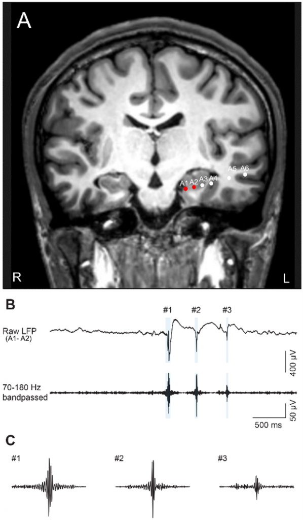
Fig. 1.
(A) An image showing preoperative magnetic resonance imaging (MRI) and postimplantation computed tomography (CT) data illustrating the electrode placement within the hippocampus. (B) Hippocampal LFPs are the bipolar potential recorded from A1–A2 (top) processed using a bandpass filter with a range of 70 to 180 Hz (bottom). The blue shaded area indicates the detected ripples. (C) Magnified views of three representative ripples.
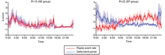
Fig. 2.
The lines show the mean and 95% confidence intervals of the Z-scored ripple event rate (red) and delta power (blue) over 24 h for patients in the NE group (left) and EP group (right).
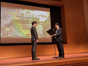
On March 8, Dr. Takamitsu Iwata received the Early Career Presentation Award at The 27th Japan Human Brain Mapping Society.
http://jhbm27.umin.ne.jp/index.html
Presentation Title: “Hippocampal sharp-wave ripples correlate with self-generated thoughts in humans”
Congratulations!

[The 27th Japan Human Brain Mapping Society]
Dates: March 7 (Fri) and 8 (Sat), 2025
Venue: Hitotsubashi Hall,National Center of Sciences Building
In November 2024, Prof. Yanagisawa became a professor at the Department of Neuroinformatics, Graduate School of Medicine, Osaka University.
We promote research activities with the goal of developing neuroinformatics to improve, restore, or alternate motor function as well as to diagnose and treat diseases and manage health.
Please find more details about our research below.
https://www.med.osaka-u.ac.jp/eng/introduction/research-5/sport/neuroinformatics-2
We appreciate your continued support.
A video byte of Dr Iwata’s Dr Iwata’s paper in Nature Communications, which was recently announced in NEWS, has been created and is available on Youtube.
For more information on Dr Iwata’s paper, please click here.
The video byte can be viewed here.YouTube LINK
Dr Takamitsu Iwata, who belonged to the Yanagisawa Lab as a graduate student until last year, has just published a Dr Iwata’s paper in the British scientific journal Nature Communications (online) on May 22, which was also published in the press on the same day.
This is the scene at the press conference. (Left: Dr Takamitsu Iwata, Right: Prof Yanagisawa)

Dr Iwata is still active in the Yanagisawa Lab as a researcher.
Congratulations, Dr Iwata!
A paper by Ryohei Fukuma, a specially-appointed lecturer in our laboratory, has been published in Communications biology.
Title of the paper: ‘Fast, accurate, and interpretable decoding of electrocorticographic signals using dynamic mode decomposition’.
A method has been developed for extracting features from multi-channel time series signals using dynamic mode decomposition, making the features easy to use in machine learning. The accuracy of brain information decoding has been improved and the computational speed is fast enough to be applied online.
Intern Yuya Ikegawa’s Dr Iwata’s paper was also published in the Journal of Neural Engineering.
Title of the paper: ‘Text and image generation from intracranial electroencephalography using an embedding space for text and images’.
Congratulations to Dr Fukuma and Ikegawa-san!
The JST CREST Brain Expression Space Interaction was featured on JST news.
Neurofeedback training was developed to change phantom hand representation without awareness of phantom limb motion or phantom limb images, successfully decreasing pain.
T. Yanagisawa, R. Fukuma, B. Seymour, M. Tanaka, O. Yamashita, K. Hosomi, H. Kishima, Y. Kamitani, Y. Saitouh, Neurofeedback Training without Explicit Phantom Hand Movements and Hand-Like Visual Feedback to Modulate Pain: A Randomized Crossover Feasibility Trial, Journal of Pain, 2022 Aug 3; S1526-5900(22)00368-6. PMID: 35932992, DOI: 10.1016/j.jpain.2022.07.009
Fukuma R, Yanagisawa T, Nishimoto S, Sugano H & Tamura K, Voluntary control 1 of semantic neural representations by imagery with conflicting visual stimulation, Communications biology, (2022) 5:214 | https://doi.org/10.1038/s42003-022-03137-x | www.nature.com/commsbio.
EurekAlert!
https://www.eurekalert.org/news-releases/946583
AlphaGalileo
https://www.alphagalileo.org/Item-Display/ItemId/218862
Using intracranial EEG, we have developed BCI, which presents some images of an imagined meaning on the screen. In addition, we have confirmed that when humans control images by their imagery while looking at a screen, brain activity corresponding to the imagined meaning is observed. This technology will be applied for reconstruction of communication function of severely paralyzed patients and novel information technology to search information based on imagery.
