


How Does the Cochlea of the Inner Ear Work?
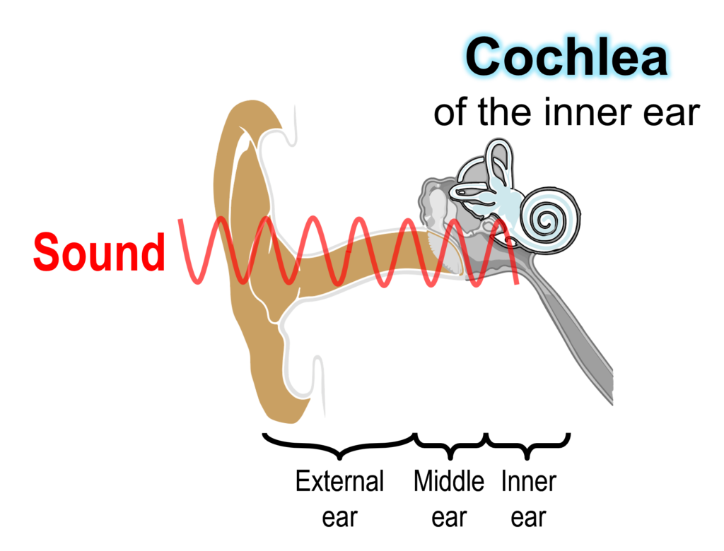
Sounds from the outside are propagated through the outer and middle ears and then reach the cochlea of the inner ear (Fig. 1).
Lower panel in Figure 2 illustrates the cross section of the cochlea. This organ has three tubular structures, which are filled with extracellular body fluids, lymph, as mentioned below. The tubules are separated by multiple cell layers.
When the cochlea is uncoiled, inside this organ a trapezoid cell layer called ‘sensory epithelium’, which converts sounds to electrical signals (upper panels in Fig. 2). Acoustic stimuli vibrate the epithelium up and down―this motion is surprisingly small of nanometer scale level. In the cochlea, the apex portion is sensitive to low pitch sounds, whereas the base portion, which is close to the entrance of the cochlea, responds to high pitch sounds. This characteristics depends on property of the sensory epithelium. This tissue is getting softer and thinner as you are approaching to the apex portion. This physical gradient plays key roles in tonotopic organization.
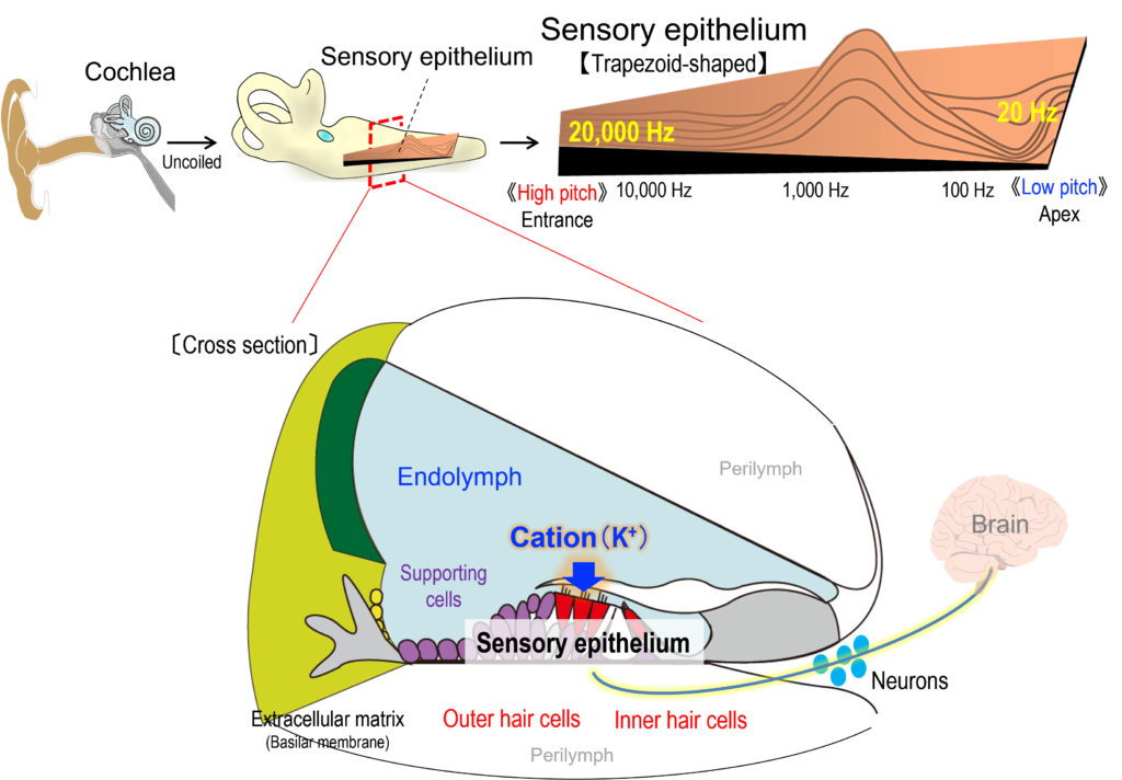
Take a look at the cross section of the cochlea (lower panel in Fig. 2). The sensory epithelium is composed of three layers, i.e. hair cell layer, supporting cell layer, and extracellular matrix layer termed ‘basilar membrane’. Sensory hair cells have hair bundle of stereocilia on the apical surface. Acoustic inputs firstly stimulate the basilar membrane and vibrates the sensory epithelium. This event deflects the stereocilia of the hair cells and further activates ‘mechanoelectrical transduction channels’ at their tips. The channel’s opening enters K+ in the lymph into the hair cells and electrically excites them. In this process, mechanical energy of sounds is transduced to electrical signals. Finally, neurotransmission occurs in the cell bodies of hair cells and acoustic information is conveyed to the brain through neural fibers.
The hair cells are categorized into two different groups; one row of inner hair cells and three rows of outer hair cells (lower panel in Fig. 2). As mentioned above, inner hair cells secrete neurotransmitters to the fibers; therefore, these cells are in charge of transmit of acoustic signals to the neuron and brain. Outer hair cells can be contracted in response to cellular electrical excitation induced by the vibrations of sensory epithelium. In other words, outer hair cells are piezoelectric elements and underlie active amplification mechanism observed in the cochlea. The cells control the sensitivity of hearing.
Get back the uncoiled sensory epithelium described in upper panel of Figure 2. Along the longitudinal axis of the epithelium, 4,000 sets of inner hair cell (one row) and outer hair cells (three rows) are beautifully arrayed. Young people who have normal hearing can perceive sounds that range from 20 Hz to 20,000 Hz (i.e., 20 kHz).
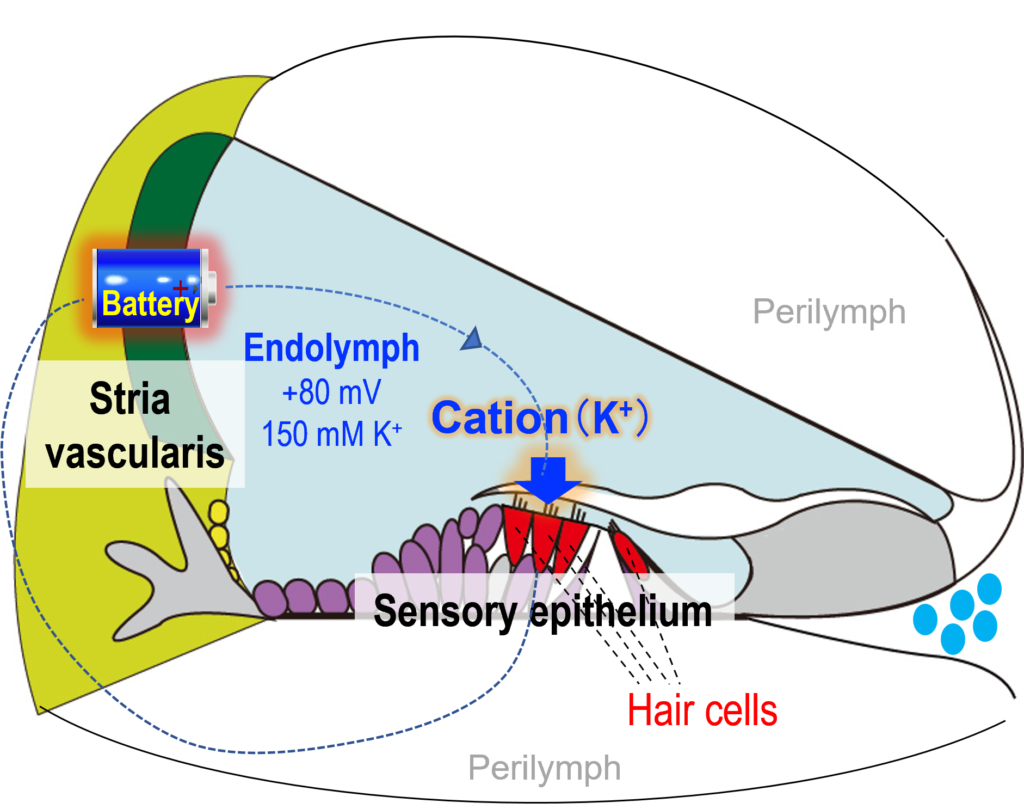
The cochlea has another element that amplifies acoustic signals. This is a biological battery in stria vascularis, which is an epithelial-like layer (Fig. 3). The battery is electrically connected to hair cells and sensitizes hearing by accelerating the K+-flux into the cells in response to mechanical stimulation. A large electromotive force measured in the battery is generated by [K+] gradient across the cell membrane between extracellular and intracellular spaces in the stria vascularis. The battery maintains a highly positive potential of +80 – +100 mV in the endolymph, where the hair bundle on the apical surface of hair cells is exposed. In addition, the endolymph exhibits high [K+] of 150 mM, low [Na+] of 2 – 5 mM, and low [Ca2+] of 20 µM. These electrochemical properties are observed only in the cochlea but not detected in any other organs. Notably, among three tubular structures of the cochlea, the center tubule is filled with the endolymph, whereas the upper and lower tubules contain the perilymph whose ion composition is similar to the regular extracellular solution such as blood plasma (i.e., 150 mM [Na+] and 5 mM [K+]) (Fig. 3).
As mentioned above, the cochlea results from assembling of machineries in the sensory epithelium and stria vascularis―orchestration of these elements achieves high sensitivity and sharp tuning observed in hearing. Our group studies the roles of each element, architecture of the network, and the relevance to auditory function and hearing loss, from cellular to organ levels by means of a variety of approaches.